Anatomy Label Major Arteries And Veins - 32 Label The Major Arteries And Veins Labels For Your Ideas : Together with teres minor muscle, teres major muscle forms the axillary space, through which several important arteries and veins pass.
byAdmin-
0
Anatomy Label Major Arteries And Veins - 32 Label The Major Arteries And Veins Labels For Your Ideas : Together with teres minor muscle, teres major muscle forms the axillary space, through which several important arteries and veins pass.. Aug 07, 2020 · the arteries were superficially labeled, primarily as they form basic anatomical landmarks. These general diagrams show the digestive system, with the major human anatomical structures labeled (mouth, tongue, oral cavity, teeth, buccal glands, throat, pharynx, oesophagus, stomach, small intestine, large intestine, liver, gall bladder and pancreas). Pulmonary arteries, and the left ventricle pumps blood into the aorta, which is the major artery in the body. Teres major is supplied primarily by the lower subscapular nerve and additionally by the thoracodorsal nerve (middle subscapular nerve). Together, heart disease and stroke, along with other cardiovascular diseases, are among the most widespread and costly health problems facing the nation today.
Together, heart disease and stroke, along with other cardiovascular diseases, are among the most widespread and costly health problems facing the nation today. For example, the brachiocephalic artery carries blood into the brachial (arm) and cephalic (head) regions. You will learn the major structures of the heart by dissecting the sheep heart. Teres major is supplied primarily by the lower subscapular nerve and additionally by the thoracodorsal nerve (middle subscapular nerve). One of its branches, the subclavian.
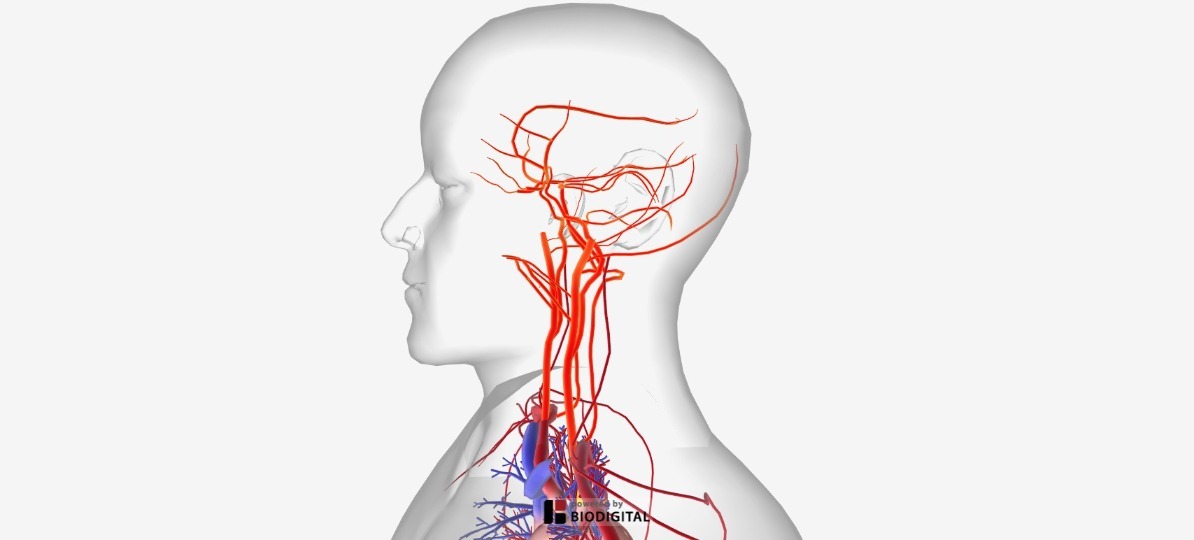
Arteries Of The Body Picture Anatomy Definition More from human.biodigital.com The major veins drain blood from the same organs and limbs that the major arteries supply. Aug 07, 2020 · the arteries were superficially labeled, primarily as they form basic anatomical landmarks. One of its branches, the subclavian. Jul 29, 2020 · there are three major types of blood vessels: These general diagrams show the digestive system, with the major human anatomical structures labeled (mouth, tongue, oral cavity, teeth, buccal glands, throat, pharynx, oesophagus, stomach, small intestine, large intestine, liver, gall bladder and pancreas). Together, heart disease and stroke, along with other cardiovascular diseases, are among the most widespread and costly health problems facing the nation today. Veins are blood vessels that bring blood high in carbon dioxide back to the heart. Correctly label the following features of the aorta and its major branches.
You will learn the major structures of the heart by dissecting the sheep heart.
While you are examining the major structures of the sheep heart, compare them with the corresponding organs of the human heart model. Correctly label the following major systemic arteries. Most deep vein blood clots occur in the lower leg or thigh. Correctly label the following major systemic veins. Heart disease is the leading cause of death in the united states.stroke is the fifth leading cause of death in the united states. You will learn the major structures of the heart by dissecting the sheep heart. Describe the anatomy of the aorta and its major branches and relate it with their functions. Together with teres minor muscle, teres major muscle forms the axillary space, through which several important arteries and veins pass. Teres major is supplied primarily by the lower subscapular nerve and additionally by the thoracodorsal nerve (middle subscapular nerve). Veins are blood vessels that bring blood high in carbon dioxide back to the heart. Jul 29, 2020 · there are three major types of blood vessels: According to the national heart and lung institute, deep vein thrombosis, or dvt, is a blood clot that forms in a vein deep in the body. Describe the basic process of hematopoiesis, where it occurs, and the significance of the pluripotent stem cell (hemocytoblast) in the process.
Most deep vein blood clots occur in the lower leg or thigh. One of its branches, the subclavian. Heart disease is the leading cause of death in the united states.stroke is the fifth leading cause of death in the united states. Blood vessels are often named after either the region of the body through which they carry blood or for nearby structures. Correctly label the following major systemic arteries.
1 from While you are examining the major structures of the sheep heart, compare them with the corresponding organs of the human heart model. Aug 07, 2020 · the arteries were superficially labeled, primarily as they form basic anatomical landmarks. The veins include the upper and lower vena cava system as well as the portal system. The major veins drain blood from the same organs and limbs that the major arteries supply. One of its branches, the subclavian. Together with teres minor muscle, teres major muscle forms the axillary space, through which several important arteries and veins pass. Blood vessels are often named after either the region of the body through which they carry blood or for nearby structures. For example, the brachiocephalic artery carries blood into the brachial (arm) and cephalic (head) regions.
Correctly label the following major systemic veins.
Aug 07, 2020 · the arteries were superficially labeled, primarily as they form basic anatomical landmarks. Together with teres minor muscle, teres major muscle forms the axillary space, through which several important arteries and veins pass. These general diagrams show the digestive system, with the major human anatomical structures labeled (mouth, tongue, oral cavity, teeth, buccal glands, throat, pharynx, oesophagus, stomach, small intestine, large intestine, liver, gall bladder and pancreas). Correctly label the following features of the aorta and its major branches. Teres major is supplied primarily by the lower subscapular nerve and additionally by the thoracodorsal nerve (middle subscapular nerve). Blood vessels are often named after either the region of the body through which they carry blood or for nearby structures. Describe the anatomy of the aorta and its major branches and relate it with their functions. Together, heart disease and stroke, along with other cardiovascular diseases, are among the most widespread and costly health problems facing the nation today. Veins are blood vessels that bring blood high in carbon dioxide back to the heart. Pulmonary arteries, and the left ventricle pumps blood into the aorta, which is the major artery in the body. Most deep vein blood clots occur in the lower leg or thigh. For example, the brachiocephalic artery carries blood into the brachial (arm) and cephalic (head) regions. According to the national heart and lung institute, deep vein thrombosis, or dvt, is a blood clot that forms in a vein deep in the body.
The veins include the upper and lower vena cava system as well as the portal system. You will learn the major structures of the heart by dissecting the sheep heart. Aug 07, 2020 · the arteries were superficially labeled, primarily as they form basic anatomical landmarks. The major veins drain blood from the same organs and limbs that the major arteries supply. Correctly label the following features of the aorta and its major branches.
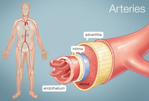
The Arteries Human Anatomy Picture Definition Conditions More from img.webmd.com For example, the brachiocephalic artery carries blood into the brachial (arm) and cephalic (head) regions. One of its branches, the subclavian. Correctly label the following major systemic arteries. Describe the basic process of hematopoiesis, where it occurs, and the significance of the pluripotent stem cell (hemocytoblast) in the process. Together with teres minor muscle, teres major muscle forms the axillary space, through which several important arteries and veins pass. The major veins drain blood from the same organs and limbs that the major arteries supply. Correctly label the following features of the aorta and its major branches. Correctly label the following major systemic veins.
Correctly label the following features of the aorta and its major branches.
Veins are blood vessels that bring blood high in carbon dioxide back to the heart. For example, the brachiocephalic artery carries blood into the brachial (arm) and cephalic (head) regions. Describe the features of blood that give it the characteristics of a connective tissue. You will learn the major structures of the heart by dissecting the sheep heart. These general diagrams show the digestive system, with the major human anatomical structures labeled (mouth, tongue, oral cavity, teeth, buccal glands, throat, pharynx, oesophagus, stomach, small intestine, large intestine, liver, gall bladder and pancreas). Together with teres minor muscle, teres major muscle forms the axillary space, through which several important arteries and veins pass. The veins include the upper and lower vena cava system as well as the portal system. Describe the basic process of hematopoiesis, where it occurs, and the significance of the pluripotent stem cell (hemocytoblast) in the process. Describe the anatomy of the aorta and its major branches and relate it with their functions. Aug 07, 2020 · the arteries were superficially labeled, primarily as they form basic anatomical landmarks. Jul 29, 2020 · there are three major types of blood vessels: Correctly label the following major systemic arteries. Heart disease is the leading cause of death in the united states.stroke is the fifth leading cause of death in the united states.